Seeing the Full Picture: Why Advanced Imaging Matters in Modern Pet Care
When our pet suddenly limps, skips a meal, or just isn’t themselves, worry sets in fast. At Omega Veterinary Group in San Mateo, California, we share that concern—and we’ve made it our mission to combine compassionate care with cutting-edge diagnostics. Central to that approach is advanced imaging: CT scans, ultrasound, digital X-rays, and MRI.
These tools allow us to see what your pet can’t say, identify serious issues early, and develop more precise treatment plans—all without surgery or guesswork.
Why Looking Inside Matters
Physical exams and bloodwork are vital, but many conditions hide deep within the body. We see firsthand how advanced imaging transforms outcomes:
- A splenic tumor spotted before it ruptures
- A complex fracture mapped in detail before orthopedic surgery
- A coughing dog diagnosed with bronchitis instead of heart failure
Each of these cases began with vague symptoms and ended with clarity, thanks to imaging.
CT Scans: Three-Dimensional Clarity
Computed Tomography (CT) scans create detailed, 3D images of bones, organs, and blood vessels by compiling thin X-ray “slices.” We recommend CT when:
- A lameness doesn’t show on standard X-rays
- A cat has chronic nasal discharge, and cancer or polyps are suspected
- A senior pet develops sudden neurological signs
CT scans require brief anesthesia to prevent motion blur, but the exposure is low and considered safe. For an accessible explanation of the process, see Why Does My Dog Need a CT Scan, Not a Simple X-Ray?.
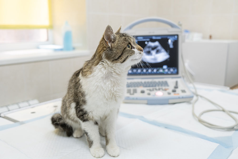
Ultrasound: Gentle, Real-Time Insight
Ultrasound uses sound waves—not radiation—to produce live images of soft tissues. It’s non-invasive, painless, and often performed with the owner nearby.
Common uses include:
- Identifying intestinal blockages in vomiting dogs
- Assessing liver blood flow in cats with weight loss
- Ruling in or out heart disease in coughing seniors
To learn more, see Small Animal Ultrasound Diagnostic Imaging – UC Davis Veterinary Medicine or Ultrasound Examination in Dogs.
Digital X-Rays: The Diagnostic Workhorse
Digital radiography remains a frontline tool for evaluating bones, lungs, and gastrointestinal structures. Results are immediate, adjustable, and easy to share with specialists. We use it to:
- Evaluate fractures, arthritis, or foreign bodies
- Detect pneumonia or tumors in the lungs
- Measure heart size and shape
Explore Small Animal X-ray Diagnostic Imaging – UC Davis Vet Med to learn more about its advantages.
MRI: Exceptional Detail When It Counts
While we don’t have an MRI unit on-site, we coordinate referrals for advanced soft-tissue studies. MRI is especially valuable for:
- Brain tumors
- Spinal cord injuries
- Cruciate ligament and meniscal damage
See Small Animal MRI Guide – Hallmarq for an in-depth look at what MRI can reveal.
When We Recommend Imaging
Imaging may be recommended when symptoms are persistent, unexplained, or inconsistent with initial exam findings. Common examples:
- Ongoing GI upset despite normal lab work
- Seizures or sudden balance problems
- A new heart murmur
- Post-trauma screening
- Unexplained weight loss
Catching conditions like adrenal tumors or liver shunts early prevents long-term damage and costly complications.
How to Prepare Your Pet—and Yourself
Before imaging:
- Fasting: 8–12 hours without food if sedation or anesthesia is needed
- Medications: Continue most, but alert us if your pet takes insulin or cardiac meds
- Bring comfort items: A familiar blanket or toy can reduce stress
On the day of the test:
- Physical exam to ensure your pet is safe for anesthesia
- IV catheter placement for fluids and medication
- 10–30 minutes in the imaging suite
- Supervised recovery, with most pets going home the same afternoon
We interpret many results immediately. Complex cases are shared with boarded specialists, and we’ll call you as soon as we have answers.
Frequently Asked Questions
Is imaging safe?
Yes. We use modern, low-dose protocols and closely monitor every patient.
Will my insurance cover it?
Most pet insurance plans reimburse advanced imaging after deductibles. We can assist with paperwork before you leave.
Can I be present?
Owners are welcome for ultrasounds when possible. For CT or X-ray, safety protocols require pets to be alone in the suite, but we reunite you immediately afterward.
How much does it cost?
Costs vary depending on the type of scan and patient size. We always provide clear estimates and discuss options upfront.
What We’re Looking For
- Bones: Micro-fractures, joint damage, signs of arthritis
- Chest: Heart enlargement, fluid around lungs, signs of cancer
- Abdomen: Liver and kidney abnormalities, masses, obstructions
- Soft tissues: Tumors, cysts, or lymph node changes invisible on physical exam
Types of Veterinary Medical Tests – Merck Veterinary Manual provides a detailed guide to what each test can detect.
When Imaging Becomes Urgent
Please seek immediate care if your pet shows:
- Rapid abdominal swelling or pale gums
- Repeated, unproductive vomiting
- Sudden collapse or paralysis
- Labored breathing or open-mouth respiration
Same-day imaging may be necessary—and lifesaving.
Long-Term Benefits of Early Imaging
Advanced diagnostics don’t just identify disease—they help us intervene earlier and treat more effectively. Whether it’s correcting a liver shunt in a growing puppy or mapping a tumor before it spreads, imaging helps us provide your pet with a longer, more comfortable life.
Our Commitment to You and Your Pet
From your first phone call to final follow-up, we aim to make your experience informed, supportive, and reassuring. Meet the professionals who make that possible on our team page, explore our full list of services, or contact us directly with any questions.
At Omega Veterinary Group, we believe that knowledge is power—and that power starts with seeing the full picture.


Leave A Comment